
Discover below the anatomical description of the 14 models available on our January 2026 test arcade:
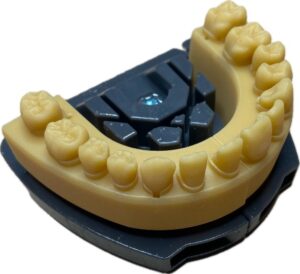
Maxillary sector 1:
- 11-01: Maxillary central incisor of a middle-aged patient with a single canal. The pulp chamber is normal and the pulp diameter gradually and evenly narrows toward the apex. The apical diameter is 15/100. The apical foramen is palatal in relation to the apex.
- 12-02-O-CAL: Maxillary lateral incisor of an elderly patient or following trauma. Absence of pulp chamber. Access cavity created. Very thin canal measuring 6/100ths.
- 13-01: Long maxillary canine of a middle-aged patient. Apical narrowing with distal and palatal curvature. The pulp section is oval and flattened in the vestibular-palatal direction. Apical diameter of 15/100.
- 14-02: Maxillary premolar with a large pulp chamber. Relatively straight palatal canal with an apical diameter of 10/100. Vestibular canal slightly curved mesially with an apical vestibular hook.
- 15-03: Second maxillary premolar with two canal entrances and a common exit (Vertucci type III). Apical diameter of 15/100.
- 16-01-REA: First maxillary molar. Relatively wide palatal canal with an apical diameter of 20/100. The distal canal has a slight apical curvature with a diameter of 10/100. The coronal portion is slightly angled. The mesial root has two independent canals. Dentin overhangs are reproduced at the entrance to these two canals. Ceramic crown reproducing the hardness of enamel, occlusal carious lesion, and reactive pulp.
- 17-01-SIMP: Simplified maxillary second molar for educational purposes, with anatomy similar to 16-01. All canals have been retouched to make the model accessible to a wider audience.
Mandibular sector 4:
- 41-01: Mandibular incisor of a young patient with a single wide canal. Root and canal curvature in the apical third in the distal and lingual direction. The apical diameter is 25/100.
- 42-03: Mandibular incisor with two canals that join in the apical third (Vertucci type III). The lingual canal is obscured by a dentinal triangle. The apical diameter is 10/100. Apical curvature in the distal direction.
- 43-01: Mandibular canine of a middle-aged patient presenting a single canal with a vestibulo-lingual flattened canal section. Apical constriction with a diameter of 15/100. An accessory canal emerges distally in the apical third.
- 44-03: First lower premolar, bifid with two separate canals in the middle third mesially and distally. Severe angulation at the entrance to each canal. The apical diameter of each canal is 10/100. Accessory canals in the apical third lingually and distally.
- 45-01: Second lower premolar of a middle-aged patient. The pulp is large with an apical diameter of 15/100. Distal curvature in the apical third.
- 46-02-P: Mandibular molar with deep occlusal caries suggestive of rapidly progressing caries in a young child. Reactive pulp for pulp capping training.
- 47-01-RTE: Mandibular molar filled with gutta-percha and zinc oxide eugenol cement. This tooth has a Weine type II canal anatomy in each root (two independent entrances, one common exit). In the mesial root, the filling stops before the junction of the two canals. In the distal root, the clinical situation suggests a failure of anesthesia and therefore insufficient preparation and filling of the most lingual canal. The second distal canal has not been prepared.
Information required to send the RIGHT-NAO® demonstration model
1 minute to confirm your shipping details and receive the template in good condition